Cutting‑Edge Optical Imaging Uncovers Subtle Activity in Blood Immune Cells
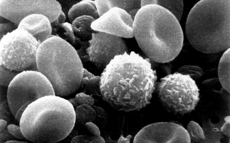
Scientists have unveiled a new optical imaging technique that can detect previously hidden functional signals in peripheral blood mononuclear cells (PBMCs), the diverse immune cells routinely isolated from a standard blood draw. The method, described in a recent pre‑print, promises to deepen insight into how these cells respond to infection, autoimmunity, and cancer without the need for invasive procedures.
PBMCs, which include lymphocytes and monocytes, are a staple of immunology research because they provide a snapshot of the body's immune status. Traditionally, researchers rely on flow cytometry, gene‑expression profiling, or cytokine assays to infer cellular activity. While powerful, those approaches often require labeling, bulk analysis, or extensive processing that can mask dynamic, single‑cell behaviors.
The newly reported technique employs advanced light‑scattering and fluorescence‑free microscopy to capture high‑resolution images of live PBMCs in real time. By analyzing subtle variations in optical density and intracellular movement, the system can differentiate between resting and activated states, even when conventional markers appear unchanged. In test samples, the imaging platform identified distinct patterns of activity in cells taken from individuals with viral infections compared with healthy donors.
According to the authors, the technology offers several practical advantages. Because it does not depend on fluorescent tags, it reduces sample preparation time and eliminates potential artifacts introduced by staining. Moreover, the label‑free nature enables repeated measurements from the same specimen, opening the door to longitudinal monitoring of disease progression or treatment response.
The implications extend beyond basic research. Clinicians could eventually use the approach to detect early immune dysregulation in autoimmune disorders or to gauge the effectiveness of immunotherapies in cancer patients. Early detection of subtle immune shifts might allow interventions before clinical symptoms become apparent, a prospect that aligns with growing interest in personalized medicine.
Nevertheless, the method remains in the validation stage. Larger studies are needed to confirm that the optical signatures consistently correlate with specific disease states across diverse populations. Integration with existing diagnostic workflows will also require standardization of imaging protocols and robust software for data interpretation.
If subsequent trials confirm its utility, the technique could become a routine component of blood‑based diagnostics, complementing current assays and offering a non‑invasive window into the immune system’s hidden activity. The research community will be watching closely as the technology moves from proof‑of‑concept toward potential clinical adoption.


Comments (0)
Be the first to comment.
Join the discussion